Experiment 2.8b – Positron Emission Tomography (PET)
The positron emission tomography is used in medicine as method to investigate physiological processes. The spatial and temporal behaviour of radioactive tracers injected into the body is observed. The method exploits the directional back-to-back correlation of the photons created in electron-positron annihilation. These are detected in coincidence by a pair of detectors.
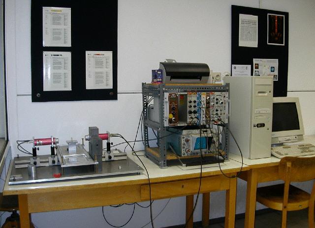
Initially, in this experiment you become familiar with measuring coincident data. The operation of BGO scintillation detectors read out by photomultiplier tubes is investigated by measuring the spectrum emitted by a 22Na source and interpreting the result. The output signals of the electronic modules used in the set-up will be explored by using an oscilloscope. Aim of the experiment is to locate radioactive sources within an enclosed container applying the PET technique. In addition, PET enables to determine the relative intensities of the sources too.